 |
Radiofrequency Radiation
|
|
Radiofrequency Radiation
|
Figures 6.25-6.30 show average SARs as a function of frequency for a few models irradiated by circularly and elliptically polarized planewaves.
Figure 6.1.
Calculated planewave average SAR in an ellipsoidal model of an average man, for the six standard
polarizations; a = 0.875 m, b = 0.195m, c = 0.098 m, V = 0.07 m3
Figure 6.2.
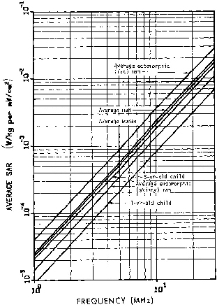
Calculated planewave average SAR in ellipsoidal models of different human-body types, EKH
polarization.
Figure 6.3.
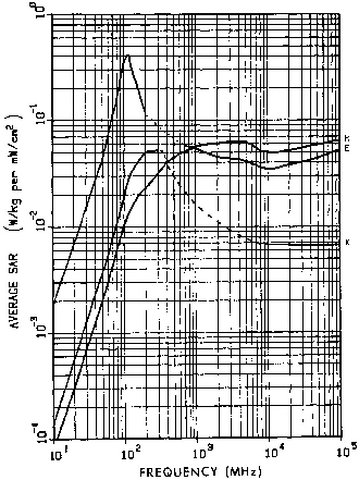
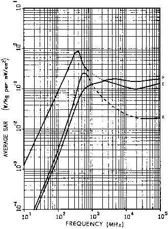
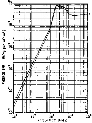
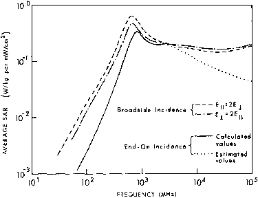
Calculated planewave average SAR in a prolate spheroidal model of an average man for three polarizations; a = 0.875 m, b = 0.138 m, V = 0.07 m3.
The dotted line is calculated from Equation 5.1; the dashed line is estimated values.
Figure 6.4.
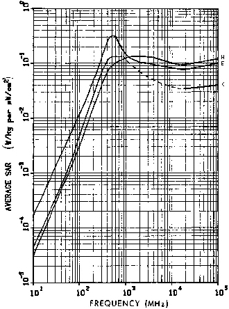
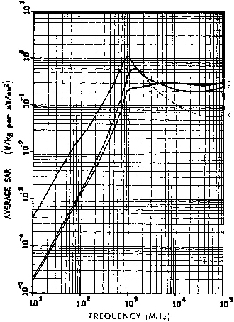
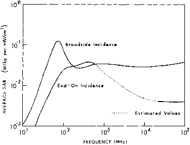
Calculated planewave average SAR in a prolate spheroidal model of an average ectomorphic (skinny) man for three polarizations; a = 0.88 m, b = 0.113 m, V = 0.04718 m3. The dotted line is calculated from Equation 5.1; the dashed line is estimated values.
Figure 6.5.
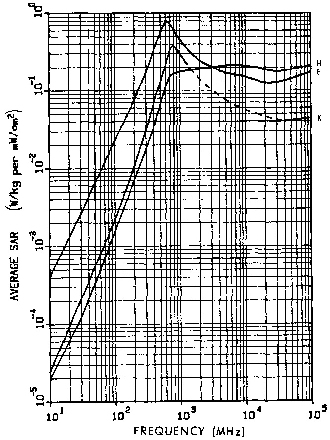
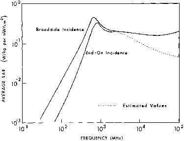
Calculated planewave average SAR in a prolate spheroidal model of an average endomorphic (fat) man for three polarizations; a = 0.88 m, b = 0.195 m, V = 0.141 m3.
The dashed line is estimated values.
Figure 6.6.
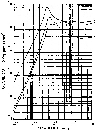
Calculated planewave average SAR in a prolate
spheroidal model of an average woman for three polarizations;
a = 0.805 m, b = 0.135 m, V = 0.06114 m3. The dotted
line is calculated from Equation 5.1; the dashed line is
estimated values.
Figure 6.7.
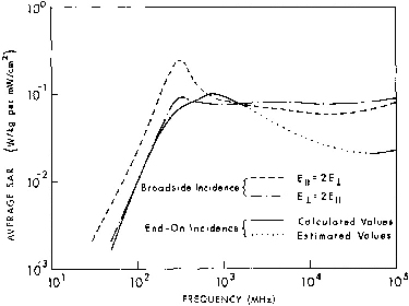
Calculated planewave average SAR in a prolate spheroidal model of a large woman, for three polarizations; a = 0.865 m, b = 0.156 m, V = 0.08845 m3. The dotted line is calculated from Equation 5.1; the dashed line is estimated values.
Figure 6.8.
Calculated planewave average SAR in a prolate spheroidal model of a 5-year-old child for three
polarizations; a = 0.56 m, b = 0.091 m, V = 0.0195 m3. The dotted line is calculated from Equation 5.1; the dashed line is estimated values.
Figure 6.9.
Calculated planewave average SAR in a prolate
spheroidal model of a 1-year-old child for three
polarizations; a = 0.37 m, b = 0.08 m, V = 0.01 m3. The
dashed line is estimated values.
Figure 6.10.
Calculated planewave average SAR in a prolate spheroidal model of a sitting rhesus monkey for three polarizations; a = 0.2 m, b = 0.0646 m, V = 3.5 x 10⁻³ m3. The dashed line is estimated values.
Figure 6.11. Calculated planewave average SAR in a prolate spheroidal model of a squirrel monkey for three polarizations a = 0.115 m, b = 0.0478 m, V = 1,1 x 10⁻³ m3. The dashed line is estimated values.
Figure 6.12. Calculated planewave average SAR in a prolate spheroidal model of a Brittany spaniel for three polarizations; a = 0.344 m, b = 0.105 m, V = 0.0159 m3. The dashed line is estimated values.
Figure 6.13. Calculated planewave average SAR in a prolate spheroidal model of a rabbit for three polarizations; a = 0.2 m, b = 0.0345 m, V = 1 x 10⁻³ m3. The dotted line is calculated from Equation 5.1; the dashed line is estimated values.
Figure 6.14. Calculated planewave average SAR in a prolate spheroidal model of a guinea pig for three polarizations; a = 0.11 m, b = 0.0355 m, V = 5.8 x 10⁻⁴ m3. The dashed line is estimated values.
Figure 6.15. Calculated planewave average SAR in a prolate spheroidal model of a small rat for three polarizations; a = 0.07 m, b = 0.0194 m, V = 1.1 x 10⁻⁴ m3. The dashed line is estimated values.
Figure 6.16. Calculated planewave average SAR in a prolate spheroidal model of a medium rat for three polarizations; a = 0.1 m, b = 0.0276 m, V = 3.2 x 10⁻⁴ m3. The dashed line is estimated values.
Figure 6.17. Calculated planewave average SAR in a prolate spheroidal model of a large rat for three polarizations; a = 0.12 m, b = 0.0322 m, V = 5.2 x 10⁻⁴ m3. The dashed line is estimated values.
Figure 6.18. Calculated planewave average SAR in a prolate spheroidal model of a medium mouse for three polarizations; a = 3.5 cm, b = 1.17 cm, V = 20 cm3. The dashed line is estimated values.
Figure 6.19. Calculated planewave average SAR in a prolate spheroidal model of a quail egg for three polarizations; a 1.5 cm, b = 1.26 cm, and V = 10 cm3
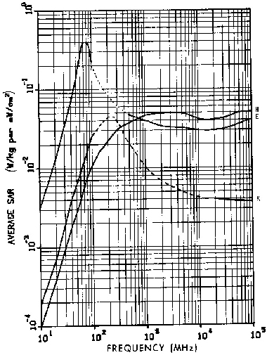
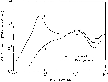
Figure 6.20. Calculated planewave average SAR in homogeneous and multilayered models of an average man for two polarizations.
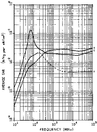
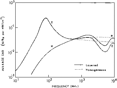
Figure 6.21. Calculated planewave average SAR in homogeneous and multilayered models of an average woman for two polarizations.
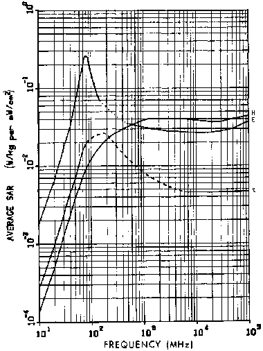
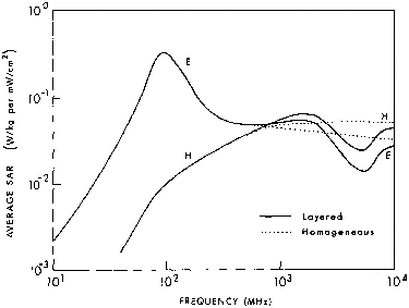
Figure 6.22. Calculated planewave average SAR in homogeneous and multilayered models of a 10-year-old child for two polarizations.
Figure 6.23. Layering resonance frequency as a function of skin and fat thickness for a skin-fat-muscle cylindrical model of man, planewave H polarization. The outer radius of the cylinder is 11.28 cm.
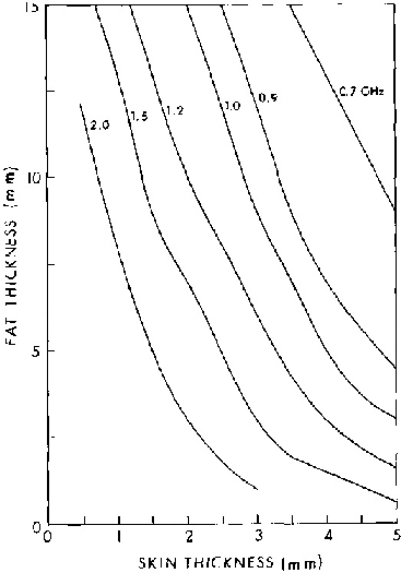
Figure 6.24. Layering resonance frequency as a function of skin and fat thickness for a skin-fat-muscle cylindrical model of man, planewave E polarization. The outer radius of the cylinder is 11.28 cm.
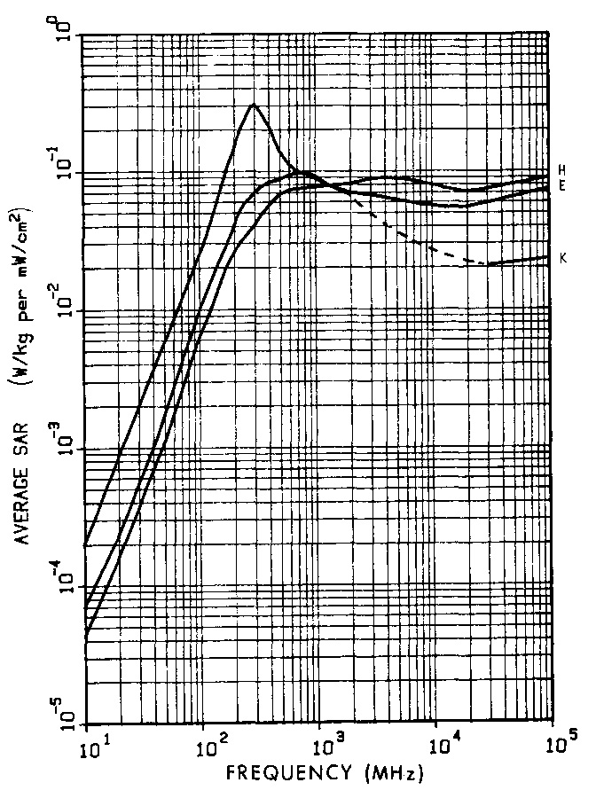
Figure 6.25. Calculated planewave average SAR in a prolate spheroidal model of an average man irradiated by a circularly polarized wave, for two orientations; a = 0.875 m, b = 0.138 m, V = 0.07 m3.
Figure 6.26. Calculated planewave average SAR in a prolate spheroidal model of a sitting rhesus monkey irradiated by a circularly polarized wave for two orientations; a = 0.2 m, b = 0.0646 m, V = 3.5 x 10⁻³ m3.
Figure 6.27. Calculated planewave average SAR in a prolate spheroidal model of a medium rat irradiated by a circularly polarized wave for two orientations; a = 0.1 m, b = 0.0276 m, V = 3.2 x 10⁻⁴ m3.
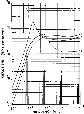
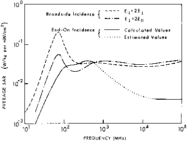
Figure 6.28. Calculated planewave average SAR in a
prolate spheroidal model of an average man irradiated by an
elliptically polarized wave, for two orientations; a = 0.875
m, b = 0.138 m, V = 0.07 m3.
Figure 6.29. Calculated planewave average SAR in a prolate spheroidal model of a sitting rhesus monkey irradiated by an elliptically polarized wave, for,two orientations; a = 0.2 m, b = 0.0646 m, V = 3.5 x 10⁻³ m3.
Figure 6.30. Calculated planewave average SAR in a prolate spheroidal model of a medium rat irradiated by an elliptically polarized wave, for two orientations; a = 0.1 m, b = 0.0276 m, V = 3.2 x 10⁻⁴ m3.
Go to Chapter 6.2
Return to Table of Contents.
Last modified: June 14, 1997
© October 1986, USAF School of Aerospace Medicine, Aerospace Medical Division (AFSC), Brooks Air Force Base, TX 78235-5301